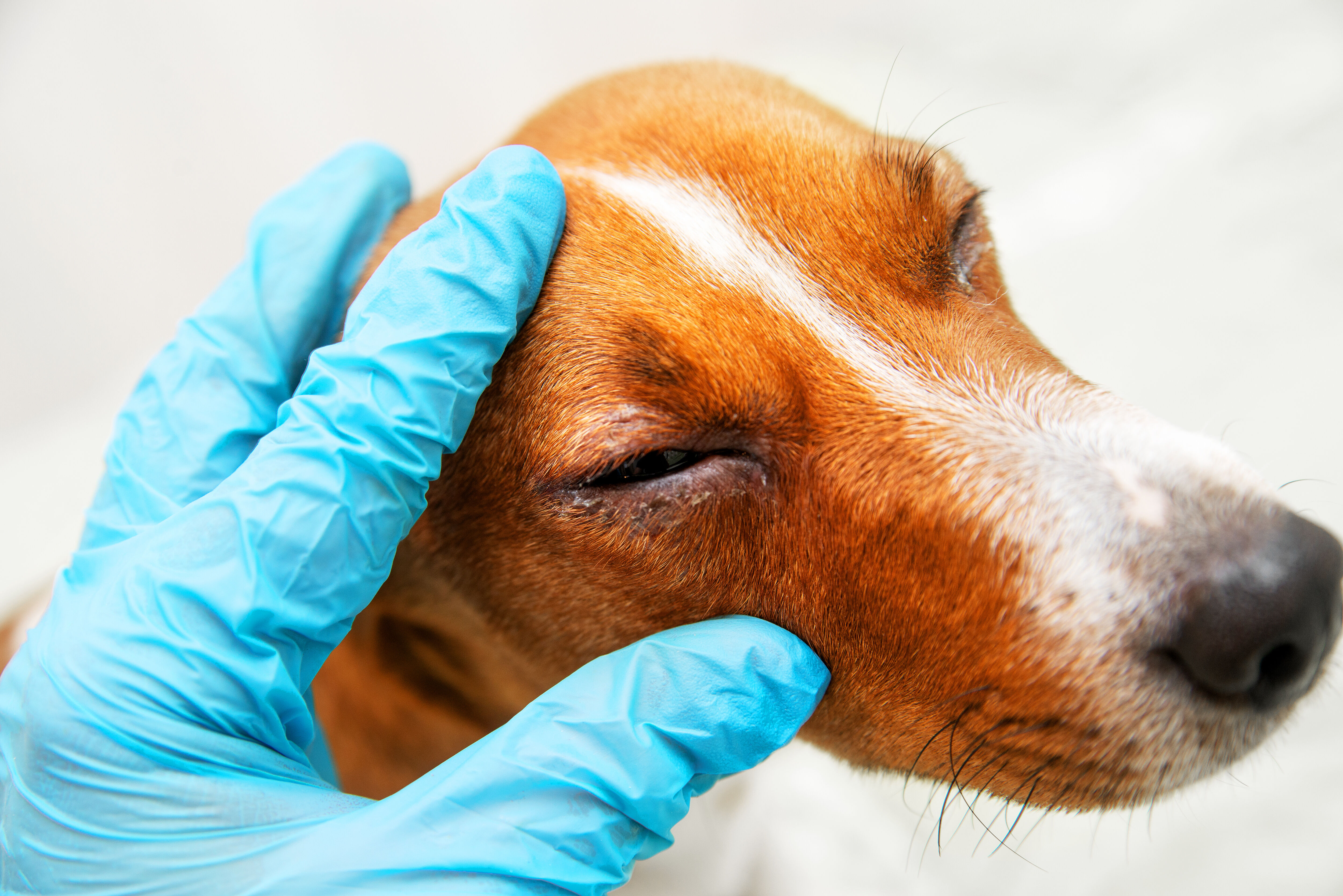
Goopy eyes it not a medical term, rather it is a complain that is used by people to describe any type of thick discharge that accumulates on the eyelids. If not washed off it becomes dry and crusts over, sometimes preventing eyelids from opening.
Why does it happen? The eyes in normal condition produce a lot of lubrication material – a tear is the liquid part that contains minerals and maintains ph, while the Meibomian glands produce a thick oil-rich part. So, if there is an intrusion, foreign body or inflammation, the eye tries to self-clean and repair and starts producing a lot of this substance. In addition, if there is an infection, white cells (Lymphocytes) come in to fight off the intruding bacteria – a pool of mucus containing these cells and bacterial cells can add to the goop.
Symptoms
It is a rare thing to have the goop without any other symptoms. Well, depending on the issue that caused the eye problem in the first place. So, symptoms may be disease-specific (when the doctor can recognize the diagnosis right away) or non-specific when the cause is not readily identifiable.
Here are some common symptoms of the goopy eyes:
Crusting of the eyelids
Inability to open the eye in the morning
Thick mucus on the eyelashes and eyelids
Tear running out of the eye without crying
Redness and irritation of the eyes
Underlying causes of goopy eyes
To understand where the goop comes from, lets take a look into the eye construction and function.
Zeis and Moll glands (ciliary glands) sit around eyelashes and produce mucin and lipids that mix with the tear to produce soft blinking without irritation of the eyelid or eye surface (conjunctiva)
A tear gland produces the tear that moisturizes the eye surface. That is what leaks out and tastes salty when we cry.
The conjunctiva of the eye consists of an epithelial layer composed of stratified squamous and stratified columnar epithelium. It is non-keratinized with interspersed goblet cells. There are also present within this epithelial layer blood vessels, fibrous tissue, lymphatic channels, melanocytes, T- and B-cell lymphocytes, Langerhans cells, and accessory lacrimal glands.
A deeper layer, the substantia propria or conjunctival submucosa, consists of superficial lymphoid and fibrous tissue. The substantia propria is a tissue layer that only exists in the conjunctiva, but not in other eye tissues. Numerous lymphocytes, mast cells, plasma cells, and neutrophils are present within this connective tissue layer.
Finally, the deepest fibrous layer contains the nerves and vessels providing innervation and blood supply to the conjunctiva. Also located within this deep layer are the glands of Krause.
Ocular surface epithelial cells produce and secrete mucins that form a hydrophilic barrier for protection and lubrication of the eye. This barrier, the glycocalyx, is formed by high molecular weight heavily glycosylated membrane-associated mucins (MAMs) that include MUC1, MUC4, and MUC16. These mucins extend into the tear film from the anterior surfaces of the conjunctiva and cornea, and, through interactions with galectin-3, prevent penetrance of pathogens into the eye.
Conjunctivitis
Conjunctivitis is an inflammation of the eye when a bacteria or virus invades (infectious), or an irritational substance gets inside (chemical and mechanical). Generally, viral and bacterial conjunctivitis are self-limiting conditions, and serious complications are rare. Because there is no specific diagnostic test to differentiate viral from bacterial conjunctivitis, most cases are treated using broad-spectrum antibiotics.
Irritants can cause chemical conjunctivitis. It is important to know which substance exactly splashed into the eye to be able to remove it promptly with a correct wash.
Foreign body in the eye
Have you ever had a tiny bug or a sand particle fly into your eye? An immediate reaction of the eye is to produce tons of tears – this will hopefully get the intruder out. But what if the piece got stuck or caused abrasion of the internal parts of the eye? Here comes that thick goop – it is the mechanism that eye switches on for a self-healing and repair.
Allergies
Allergy to environmental proteins can affect the eye and eyelid. Allergic conjunctivitis is a very common disease of children and adults. Sensitization to a protein happens before the symptoms start – a normal environmental airborn particles such as cat hair, dog hair, particles of a dried-out insects or tiny dust mites may trigger an abnormal response from the immune system. Instead of simple recognition and tolerance, sometimes there is an error, and the cells start attacking that protein causing a cascade of allergic inflammation.
After this allergic response is written in the core memory immune system, any time that protein comes in contact the symptoms start. It is not known why one person will develop nose congestion while another develops allergic conjunctivitis.
Irritant contact reactions can happen with many chemicals used commonly in the beauty products, creams or household cleaning.
Reactions to contact lens can develop with continuous wear, or using solutions that cause personal sensitivity.
Blocked tear duct
Most of the time blocked tear duct is an inherited problem that is caused by a narrow bone passage of the tear duct. The tear duct travels from the lower eyelid through the cheek bone to the internal cavity of the nose. Any constriction of that canal will cause the tear to accumulate inside the eyelid compartment and overflow over the eyelid. A significant blockage also prevents normal clearing of the eye and can lead to the inflammation and infection.
Stye
A stye, also known as a hordeolum, is a common problem involving the eye seen in both primary and urgent care setting. It is a painful, acute infectious process of the upper or lower eyelid. Classically a hordeolum appears as a small pustule along the margin of the eyelid and can be differentiated from a chalazion which tends to involve less of an inflammatory response and follows a more chronic course. An acute bacterial infection of the eyelid margin, 90% to 95% of cases of hordeolum are due to Staphylococcus aureus with Staphylococcus epidermidis being the second most common cause. An external hordeolum represents a localized abscess formation of the follicle of an eyelash whereas an internal hordeolum is an acute bacterial infection of the meibomian glands of the eyelid.
The meibomian glands are modified sebaceous glands that are found in the tarsal plate of the eyelids. They produce an oily layer on the surface of the eye that helps to maintain proper lubrication of the eye. When a meibomian gland becomes acutely infected, it results in an internal hordeolum. Due to its deeper position within the eyelid, internal hordeola have a less defined appearance than external hordeolum.
Dry eye syndrome
Dry eye is a syndrome that annoys many people worldwide. There are also many reasons that can cause it – it can be a symptom of another disease or it can happen because some eye glands are not functioning well.
A healthy ocular surface microenvironment, especially a stable tear film, is essential to preserve the smooth optical surface, epithelial cell health, ocular comfort, provide protection from environmental and microbial insults. Interconnection between the ocular surface tissues and secretory glands through the central nervous and endocrine system directs production of the tear film, has evolved a complex network to maintain ocular surface microenvironment homeostasis, especially tear film stability.
If any of the components do not work properly it can cause underproduction of lubricant and dry eyes. Dry eye syndrome can lead to many problems including bacterial infections of the eye and scarring of the eye and eyelid structures.
Keratitis
Infectious keratitis is a major global cause of visual impairment and blindness, often affecting marginalized populations. Topical antibiotics remain the best treatment for bacterial keratitis, and a recent review found all commonly prescribed topical antibiotics to be equally effective. However, outcomes remain poor secondary to corneal melting, scarring, and perforation. Viral keratitis differs from bacterial and fungal cases in that it is often recurrent and is common in developed countries.
Use of orthokeratology lenses is generally safe, but cases of associated infectious keratitis may have a higher incidence of virulent organisms such as Pseudomonas, Acanthamoeba, and antibacterial-resistant strains of Staphylococcus, partially due to the required overnight use of these lenses.
Trachoma
Trachoma is the most common infectious cause of blindness. Repeated episodes of infection with Chlamydia trachomatis in childhood lead to severe conjunctival inflammation, scarring, and potentially blinding inturned eyelashes (trichiasis or entropion) in later life. Trachoma occurs in resource-poor areas with inadequate hygiene, where children with unclean faces share infected ocular secretions.
Entropion
Entropion is a common eyelid malposition in which the margin turns inward against the globe. If untreated, this condition can cause irritative symptoms like ocular discomfort, corneal abrasion, microbial keratitis, corneal vascularization, and visual loss.
Goopy eyes in children
In children the main concerning symptoms that come with the eye discharge are:
Red eye
Burning or itching.
Photophobia
Headache
Irritability
Eye swelling
Inability to open the eye
Visual blur
This is an immediate concern of the parents that are likely to bring children straight to the urgent care.
Conjunctivitis is the most common cause of red eye. Other common causes include blepharitis, corneal abrasion, foreign body, subconjunctival hemorrhage, keratitis, iritis, glaucoma, chemical burn, and scleritis.
What does the color of my discharge mean?
There are three main colors of the discharge:
Pale while
Yellow
Green
What exactly these colors tell you? Well, any doctor will tell you that the colors of the discharge can be suggestive of the infection or another process, but there are exclusions and confusions. The rule of thumb – green goop means bacterial infection, yellow and white means everything else including foreign body, viral infection, trauma and allergy. Fungal infection may come in various colors from pale to green, so this one is difficult to tell.
Diagnosis
The goopy eye can be a one-day nuisance, or a continuous problem that is resistant to the treatments. Which is true for a diagnosis – a primary care provider can be right on top of it and cure the issue within a few days, or there may be many unsuccessful visits to various specialists trying to get to the bottom of it.
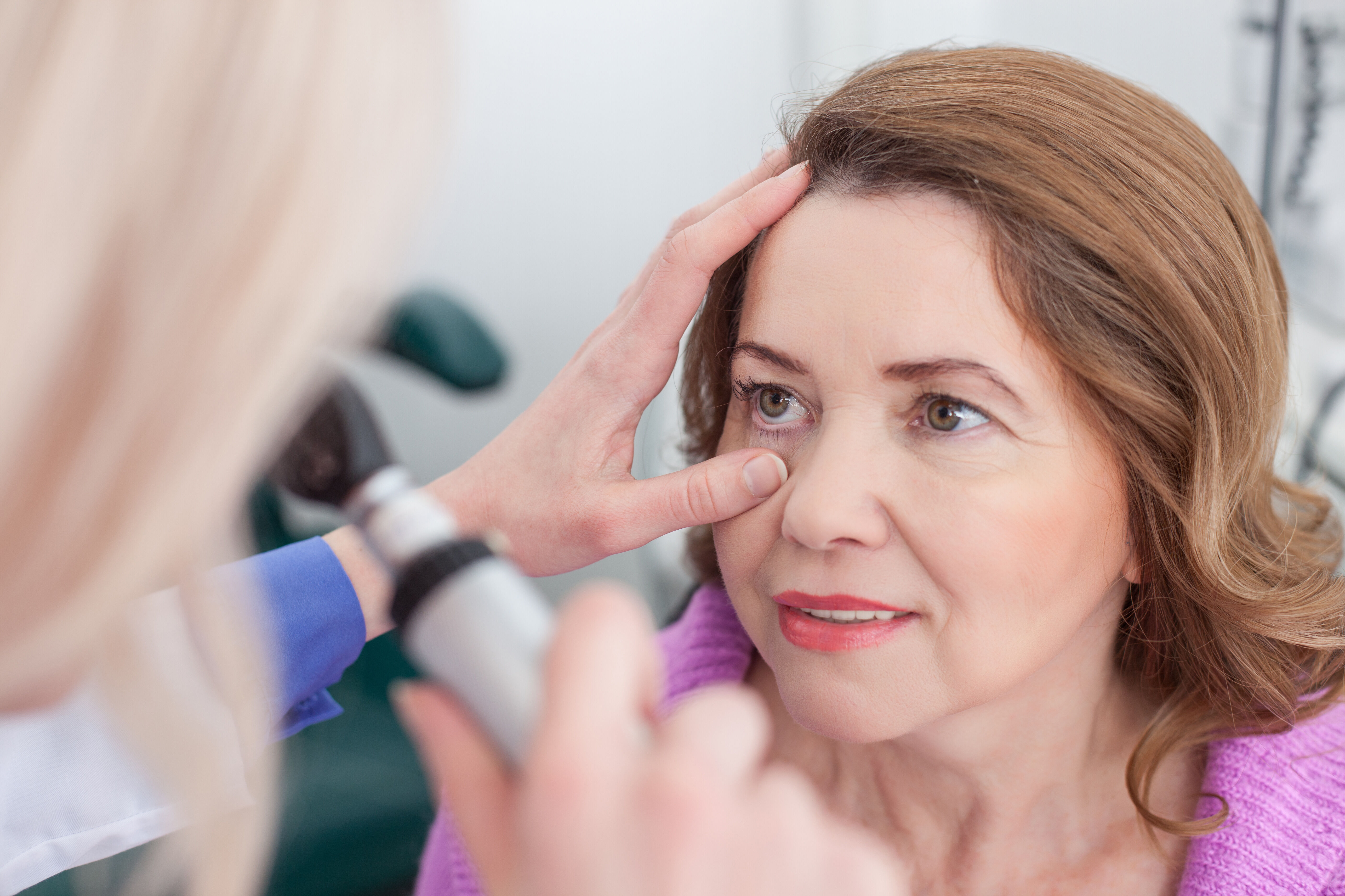
When to see a doctor?
If the discharge is persistent or comes with the variety of the symptoms – time to see a doctor. Start with the primary care – goopy eyes is a very common problem that is well-recognized by all pediatricians and internists. It is wise to see an optometrist or ophthalmologist when:
the goopy eye is not relieved with the treatment recommended by a primary care doctor;
topical steroids or other significant treatments with potential side effects were recommended;
Treatment is really dependent on the cause of the discharge. So, you can start with self-treatment if the issue does not seem to be significant. But if there is any concern it is best to see a professional. After all – it is your vision that is at stock.
Eye bacterial infections are treated with topical drops, gel or cream containing antibiotics
Viral infections need only support measures, if last less then a week. Pain and discomfort can be helped with pain killers and topical pain relief eye drops.
Foreign body, if did not wash out immediately must be removed by a professional. It is best to close the eye with the patch so the piece will not continue to damage the eye. an ophthalmologist is skilled in foreign body removal, and can examine the eye for scratches. Further treatment may be needed to prevent infection and heal the damage.
Allergic condition is treated best by an allergist if you could not identify and remove the cause. As the eye can get involved in many allergic reactions, it is best to try removing everything allergenic first and see if the condition improved.
At-home checklist
In case of the allergy there are couple of very important measures you can do:
Change the detergent. A powder from any type of detergent (even Free and Clear) can be very irritational
For women – remove all cosmetics that is used close to the eyes
Reduce dust mites
Check for new soaps, shampoos and creams
Check for any new eye drops or lens solution
Reduce the fur if you have domestic animals, note if your eyes are more itchy after you pet a cat or a dog.
Note if your symptoms are more common indoors or outdoors.
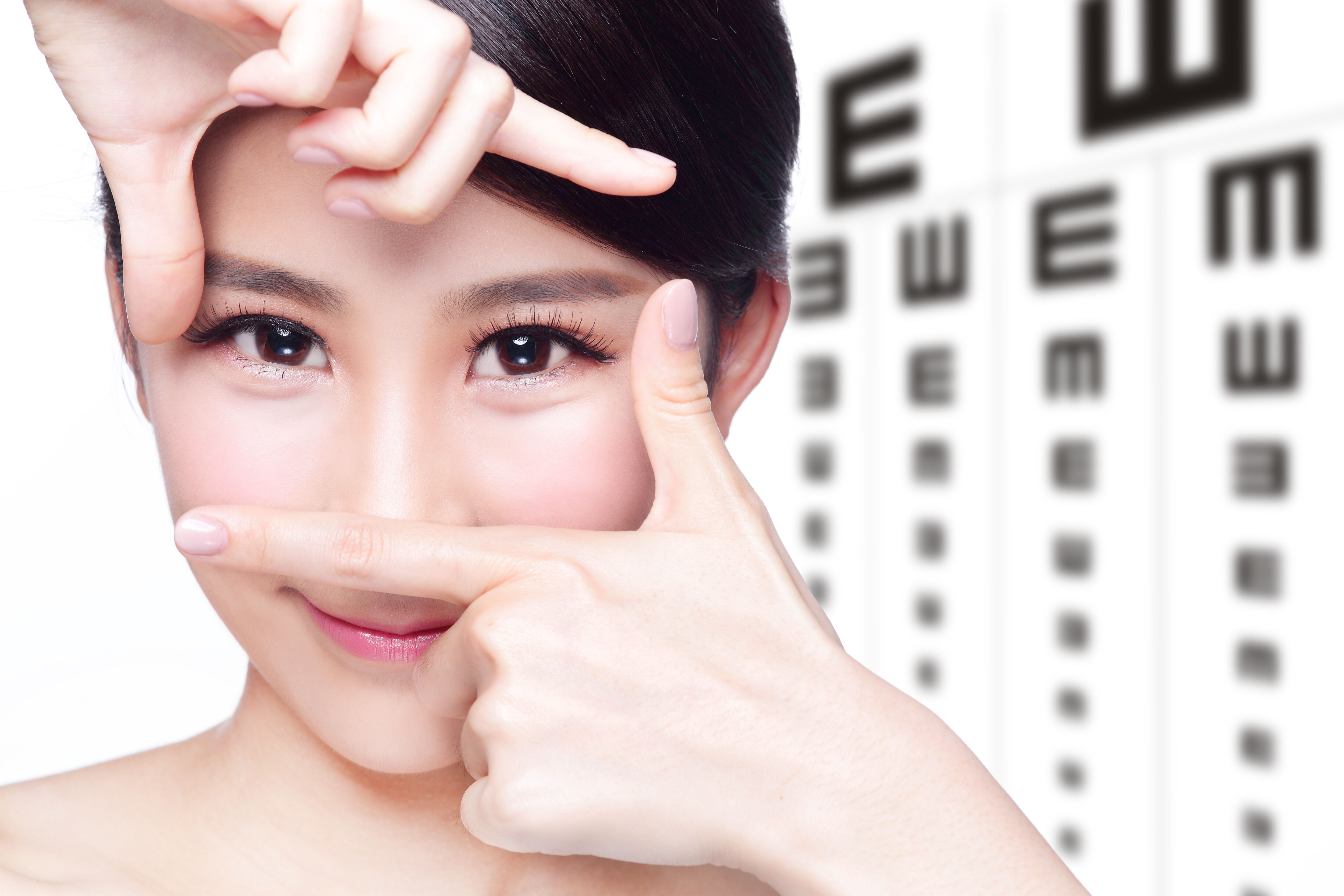
Prevention
The eyes just like all other parts of the body need to be cared for. Daily routine should include washing the eyes with water at least once a day. Good hygiene can prevent many infectious and contact problems. Teach kids not to rub the eyes and wash hands frequently – most of the bacterial and viral infections are transmitted through dirty hands and rubbing eyes. Use only baby shampoos for the babies and kids that are tear-free. It is important to use the minimal amount of cosmetics that has irritating chemicals, and always wash off mascara and eyeliner off at night. Eyes are very sensitive and it is wise to disturb their internal health and self-cleaning mechanisms.