Giant Papillary Conjunctivitis
- April 9, 2022
- by NATALIYA KUSHNIR
- Articles
What is giant papillary conjunctivitis?

Giant papillary conjunctivitis (GPC) is a syndrome found frequently as a complication of contact lenses. Many variables can affect the onset and severity of the presenting signs and symptoms. Rigid gas permeable contact lenses appear to result in less severe signs and symptoms, with a longer time before the development of giant papillary conjunctivitis. Nonionic, low-water-content soft contact lenses tend to produce less severe signs and symptoms than ionic, low-water-content soft contact lenses.
Enzymatic treatment appears to lessen the severity of signs and symptoms. The association of an allergy appears to play a role in the onset of the severity of the signs and symptoms but does not appear to affect the final ability of the individual to wear contact lenses.

Here is an ophthalmology definition of the GPC:
It is an allergen exposure and subsequent response secondary to the ocular foreign body either harboring allergens on its surface or injuring ocular structures which facilitate allergen infiltration. It can be seen with many different ocular foreign bodies (e.g., contact lenses, prostheses, cyanoacrylate glue, and sutures).
Symptoms
GPC is seen most commonly in teens and young adults, most likely because of a temporal relationship with contact lens use. Most commonly seen in conjunction with soft contact lens use and is present in approximately 5% of that population. Average onset is one to two years after starting soft contact lenses but varies widely with other ocular foreign bodies. Contact lens wearers with giant papillary conjunctivitis report a variety of symptoms, including:
- decreased lens tolerance,
- increased lens awareness,
- excessive lens movement,
- increased mucus production associated with ocular irritation,
- redness, burning, and itching.
In addition, very frequently there are all other allergic symptoms such as sneezing, congestion, runny nose, cough and postnasal drip.
Stages of GPC
Patients almost invariably have coated contact lenses and usually complain of mucus in the inner canthus on awakening in the morning. Examination of the upper tarsal conjunctiva reveals inflammation and the presence of papules, which, in the full-blown syndrome, are larger than one third of a millimeter. In the early stages, the symptoms of GPC may precede the signs.
It was suggested to classify GPC in four stages which are more important for an ophthalmologist then for allergist:
- Stage 1 symptoms consist of minimal mucus discharge in the morning, occasional itching after lens removal, and occasional coating of the contact lens. Examination of the tarsal conjunctiva may reveal a normal appearance with minimal to mild hyperemia
- Stage 2 is characterized by increased mucus with associated itching, increased lens awareness, coated contact lenses, and mild blurring of vision. Patients state that these symptoms begin a few hours after insertion of their contact lenses. These complaints may limit or decrease a patient's ability to wear the lenses. Slit-lamp examination of the upper tarsal conjunctiva reveals mild injection with slight thickening of the conjunctiva. There is mild erythema of the conjunctival plate. These changes in the tarsal conjunctival plate tend to obscure superficial vessels, although the deeper vessels usually remain visible. Papules of variable size have formed; some will be 0.3 mm or larger. One can also see coalescence of the papules, with several adjacent papules elevated as a result of the thickening of the underlying tissue. These early changes may be difficult to detect by white light. However, instillation of fluorescein and examination under cobalt illumination make them clear.
- In stage 3, itching and mucus formation have again increased. Lens coating is now frequent. Patients find it extremely difficult to keep their lenses clean. Lens awareness, especially with excessive lens movement on each blink, results in fluctuating blurred vision. Lens wearing time is substantially reduced. Examination of the upper tarsal conjunctiva shows marked injection and thickening, with further obscuration of the normal vascular pattern. Papules on the tarsus will have increased in size and number an(d become elevated.
- By stage 4, patients usually have complete lens intolerance. They note discomfort shortly after insertion of their lenses, which quickly become coated and cloudy. In addition, excessive lens movement and decentration occur. Mucus secretion is quite noticeable and may be severe enough that the patient's eyelids stick together in the morning. Papules on the upper tarsus are larger and may be flattened at their apices. As in stage 3, there is often fluorescein staining of the papule apices. Although these stages have been described to illustrate the progression of both the signs and symptoms of GPC, individual variation is common.

Some patients have severe symptoms with early tarsal changes; others are asymptomatic but, on examination, are found to have impressive inflammation and papillary reaction of the upper tarsal conjunctiva.
Possible causes

GPC is usually bilateral, but in approximately 10% of patients it is either unilateral or markedly asymmetric. In approximately half of these cases, the ophthalmologist can often find obvious reasons, such as asymmetric coating, spoiled lenses, or poor fit, to explain the disparity between the manifestation of the disease in two eyes; in the other half, no specific cause can be found.
Drs Kari and Haahtela reported that seasonal allergy is a risk factor for wearers of soft contact lenses. They also found that atopic contact lens wearers had greater problems with their lenses than did nonatopic individuals. They concluded that being atopic increases the risk of ocular problems by a factor of 5.
When individual categories of allergies were evaluated, significantly more GPC patients than control subjects reported allergic reactions to contact lens solutions (thimerosal).
There is considerable overlap with atopic conjunctivitis (vernal and atopic keratoconjunctivitis) as well as giant papillary conjunctivitis in both treatment and certain aspects of pathophysiology. As such, these are all considered to be ocular allergies.
Differential diagnosis with other eye allergic conditions is made after an ophthalmological examination:
Vernal keratoconjunctivitis (VKC)
Symptoms are usually most severe in the spring and include thick mucus discharge, pain, photophobia, and blurred vision. Patients will also often complain of foreign body sensation. On examination, corneal ulcers and conjunctival infiltrates can sometimes be found. Giant papillae on the tarsal conjunctiva are universally seen on examination.
Vernal Keratoconjunctivitis will show an accumulation of eosinophils, mast cells, and proliferation of fibroblasts. Biochemical stains will reveal the presence of proteases, chymase, and tryptase. The substantia propria is thickened due to collagen deposition. Both T and B lymphocytes are present which release IgE and IgG. The overall picture resembles both type 1 and type IV hypersensitivity reaction.
Atopic keratoconjunctivitis (AKC)
Symptoms are usually perennial and include pain, blurry vision, photophobia, and foreign body sensation. Examination reveals findings similar to simple allergic conjunctivitis with the addition of chronic inflammatory changes to the ocular surface (corneal scarring and neovascularization) and varied changes to the eyelids (lower lid more commonly) and peri-orbital skin that range from mild atopy to lichenification.
Atopic keratoconjunctivitis will also show a slight increase in eosinophils and mast cells. Tears will contain high levels of IgE. The picture resembles Type I hypersensitivity.
Secondary GPC
Atopy is more common in GPC patients than in other in individuals. By using an IgE serum level of 50 kU/l or greater to separate atopic from non-atopic patients, Buckley and colleagues found that the prevalence of atopy in GPC patients was 44%, compared with only 20% in the general population. Most patients who develop GPC report allergies to contact lens solutions, medications, molds, pollens, or animals.

Diagnosis
Careful questioning is needed to from patients to find the right diagnosis. Sometimes you will need to see couple of specialists:
At the ophthalmologist office
When the changes are seen but patients do not complain about it, it is because they may think it is normal to have an irritation.
An eye doctor will examine the inner tarsal conjunctiva to help arrive at the correct diagnosis. The normal tarsal conjunctiva, as examined by slitlamp biomicroscopy, shows a vascular arcade that appears as fine vessels radiating perpendicular to the tarsal margin. The normal conjunctival surface has a moist, pink color. GPC will have a very distinct appearance.

At the allergist office
It is important to find out what the allergen is and how to avoid it. To begin with, you will have an allergy skin prick test that can tell you for sure if it is the grass or the cat causing all the problems. If you have a clear answer, then you will discuss the possible treatment with the allergist. You will also learn about elimination and prevention measures.

Treatment
First item of management is to remove the mechanical irritant which is most commonly a contact lens. Patients should be educated and begin the same general allergic eye care used in other subtypes of ocular allergy (avoid rubbing eyes, use artificial tears and cool compresses, avoid allergen exposure). Initial pharmacotherapy is similar to other ocular allergies, for example, topical antihistamine or combination antihistamine and mast cell stabilizing drops.
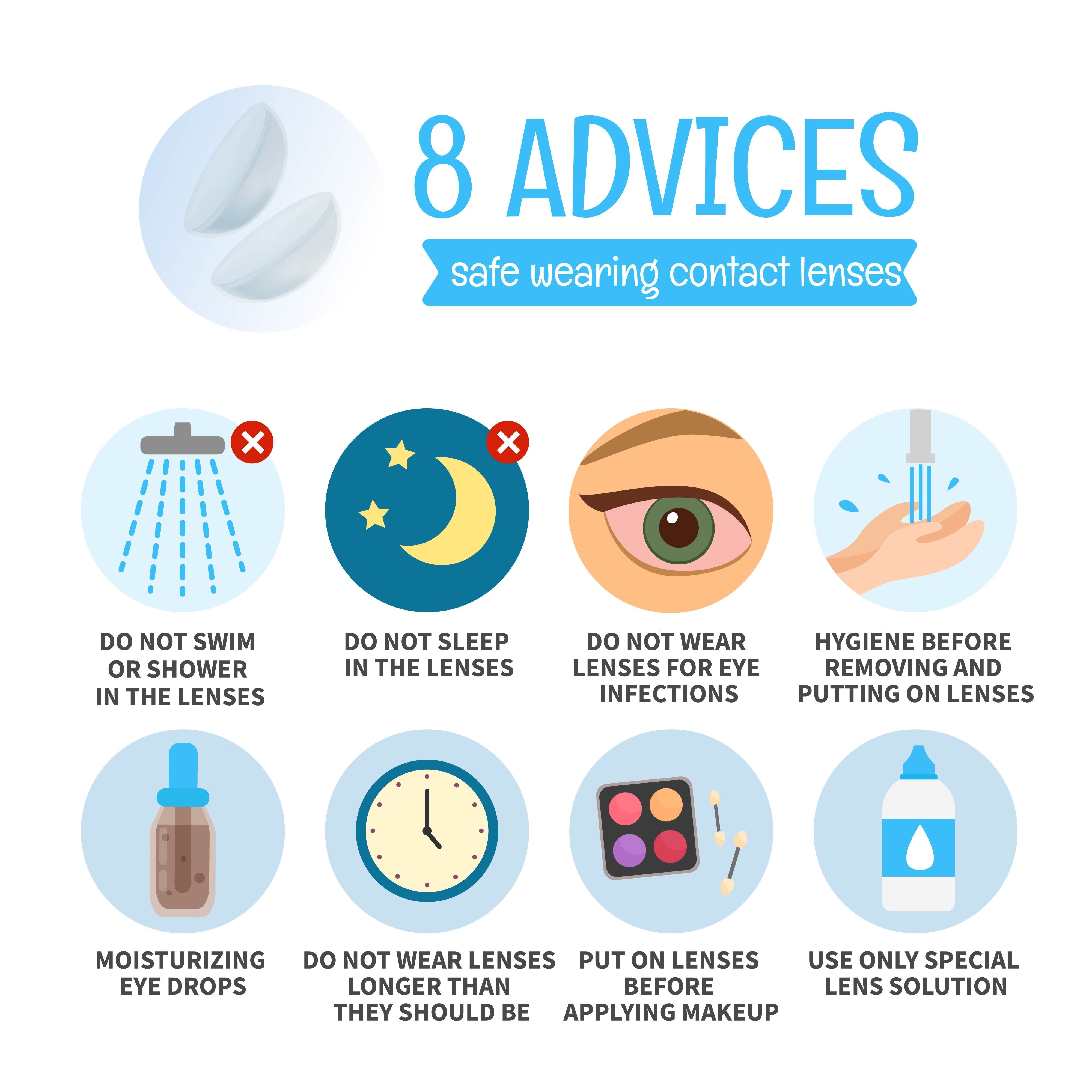
Practice proper lens care

A regimen consisting of the use of a daily cleaner, enzymatic treatment once or twice a week, and hydrogen peroxide disinfection, as well as replacement of patients' contact lenses with new ones of the same type, allowed more than a half of GPC patients to continue wearing lenses satisfactorily for an average of 15.2 months in a published study.
Change the type or design of your lenses
Use of the same care regimen, but changing to a different type of soft contact lens, enabled 77% of patients to wear their lenses for an average of 11.9 months without any significant problems.
Stop wearing contacts temporarily
Discontinuing contacts and switching to glasses for at least 2 weeks usually significantly improves symptoms. As the conjunctiva heals you may return to wearing lens without further problems.
Use prescribed eye drops
Lustein and coworkers, in a study of 47 patients with GPC, evaluated the efficacy of three treatment options: topical Opticroin 4% (sodium cromolyn) with continued lens wear; Opticrom combined with temporary (discontinuation of lens wear; and temporary discontinuation of lens wear alone. Of the 22 patients whose symptoms totally resolved, 16 were treated with Opticrom and temporary lens discontinuation.
Treating primary GPC
Treating primary GPS is always a process involving both a patient and a doctor. It is important for an ophthalmologist to give appropriate recommendations and prescribe the best possible lenses. But it is crucial for a patient to be compliant with all eyecare recommendations.
Possible complications and when to see your doctor
The long-term outcomes of PAC and SAC are good, but a significant number of people are left with eye discomfort and poor ocular cosmetic. Some individuals develop recurrences leading to conjunctivo-chalasis which is secondary to ongoing limbal conjunctival chemosis. Other complications that can occur in VKC and AKC include corneal opacification and ulcer formation. Some patients may develop lid involvement leading to difficulty in wearing contact lenses.
Here are the reasons to see a doctor:
- Treatment you have tried does not work
- You have problems with vision
- The itching and pain is increasing and unbearable
- You cannot open the eye
- You want to know the actual allergen that is causing the problem
Prevention
The main prevention is the treatment of seasonal or indoor allergy. You need to figure out what was the problem in the beginning, and eliminate it to prevent GPC. And also you need to follow all the eye doctor advices on the lens care and eyecare.
